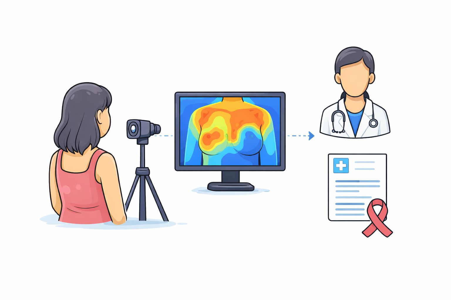
In most hospitals, diagnosis begins with an image. A chest X-ray. A CT scan. A scan that captures what the human eye cannot see directly.
But the process that follows is slower than it appears. Images are taken quickly, but interpretation depends on trained radiologists. In many parts of the world, there are simply not enough of them. Reports get delayed. Critical conditions are sometimes missed or detected late.
Qure.ai is built around this bottleneck. It does not replace medical imaging. It focuses on what happens after the image is taken—how quickly and accurately it is understood.
The origin
Qure.ai was founded in 2016 by Prashant Warier and Pooja Rao, with roots in Fractal Analytics, a company known for its work in data science and AI.
The founding idea was straightforward. Medical imaging generates large volumes of data, but extracting insights from that data depends heavily on human interpretation. If machine learning models could be trained to read these images, they could support doctors and reduce delays.
The early focus was on chest X-rays, particularly for tuberculosis, a disease where early detection is critical and screening volumes are high. From there, the scope expanded.
What Qure.ai does
Qure.ai builds AI systems that analyse medical images and generate structured outputs. The company’s core products are tied to specific imaging types.
There is qXR, which reads chest X-rays. There is qER, which analyses head CT scans, especially for stroke and brain injuries. There is qCT, which works on chest CT scans for lung conditions.
These systems are trained on large datasets of medical images. They learn patterns associated with different diseases—such as lung nodules, fractures, bleeding in the brain, or signs of tuberculosis.
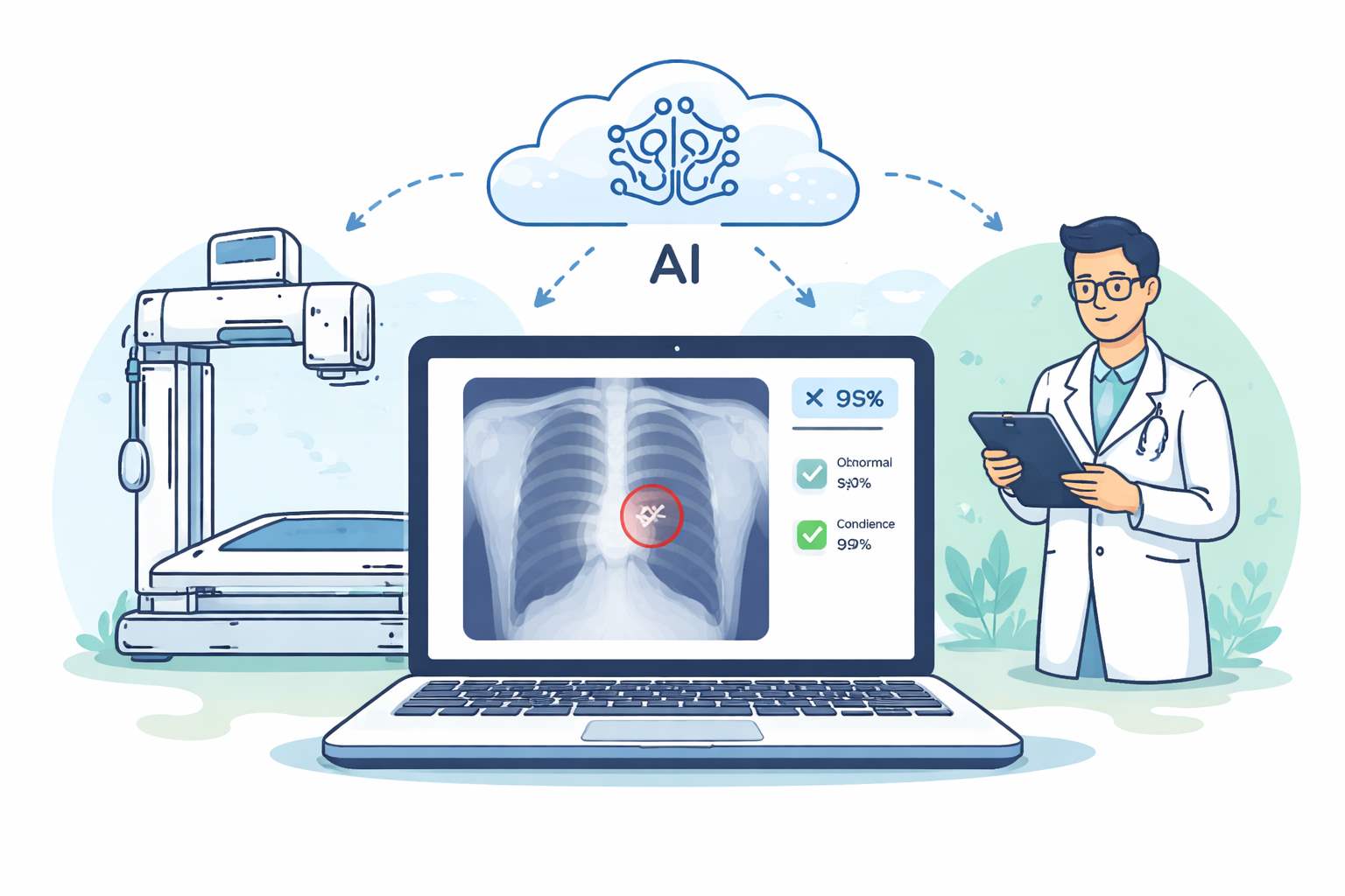
When a new image is uploaded, the system processes it and highlights areas of concern. It does not simply say “normal” or “abnormal.” It marks regions, assigns probabilities, and generates a structured report that a doctor can review. In practical terms, it acts as a second set of eyes.
How the system works
The workflow is built to fit into existing hospital systems. An image is captured using standard equipment. That image is then processed by Qure.ai’s software, either through cloud systems or local integration. The AI model analyses the image and produces outputs within seconds.
These outputs include:
highlighted regions
probability scores for conditions
prioritisation flags for urgent cases
For example, if a CT scan shows signs of a brain bleed, the system flags it as urgent so that it moves ahead in the reporting queue. This is important because in many hospitals, radiologists work through large backlogs. Prioritisation changes outcomes.
What changes in real settings
Before systems like Qure.ai, imaging workflows are sequential. Images are captured → added to a queue → reviewed when a radiologist is available. After deployment, the workflow becomes partially parallel. Images are analysed immediately → urgent cases are flagged → radiologists focus attention where it matters most. This reduces delays, especially in high-volume settings.
In stroke care, for example, minutes matter. Faster detection can directly influence treatment decisions.
In tuberculosis screening programs, large volumes of X-rays can be triaged automatically, allowing human experts to focus on confirmed or high-risk cases.
Scale and deployment
Qure.ai has scaled across multiple geographies. Its solutions are deployed in more than 90 countries and across over 3,000 healthcare sites.
In broader estimates, its systems are used in thousands of hospitals globally and have reached millions of patients annually. The company has also built strong partnerships with global healthcare organisations, governments, and pharmaceutical companies.
Funding and growth
Qure.ai has raised significant capital over multiple rounds. In 2022, it raised around $40 million from investors including Novo Holdings and HealthQuad. In 2024, it raised an additional $65 million in a Series D round led by Lightspeed and 360 ONE Asset. Overall funding has crossed $100 million, with backing from a mix of healthcare and technology investors.
The company has used this capital to expand globally, invest in new AI models, and integrate more deeply with healthcare systems.
Pilots and real-world performance
Qure.ai’s systems have been validated across multiple clinical settings.
In tuberculosis screening programs, its AI tools are used to analyse chest X-rays at scale, helping identify potential cases quickly.
In emergency care, its head CT analysis tools help detect strokes and brain injuries, enabling faster triage.
Performance metrics are often evaluated in terms of sensitivity and specificity—how accurately the system detects conditions.
Product evolution
Beyond core imaging tools, Qure.ai has expanded into workflow support. One example is AIRA, an AI-powered co-pilot designed to assist frontline healthcare workers by guiding data collection and clinical workflows.
The company is also working on integrating AI with portable imaging systems, including ultrasound, to extend diagnostics beyond hospital settings.
What makes the approach different
Qure.ai’s differentiation lies in how it combines clinical focus with scalable technology. It is not a general-purpose AI platform. Each product is designed for a specific clinical use case. It integrates directly into hospital workflows rather than requiring new systems. It focuses on high-volume, high-impact conditions like tuberculosis, lung cancer, and stroke. It is also built for varied environments, including settings with limited radiology infrastructure.
The global context
Qure.ai operates in the broader space of AI-driven medical imaging. Globally, this is a rapidly growing field. Healthcare systems are under pressure due to rising patient volumes and limited specialist availability.
AI tools are increasingly being used to assist in diagnosis, triage, and workflow management. The market is expected to grow significantly over the next decade, driven by both technological advances and healthcare demand.
Multiple companies are working in this space, but approaches vary. Some focus on specific diseases. Others build broader imaging platforms. The differentiator often lies in clinical validation and real-world deployment.
- Our correspondent