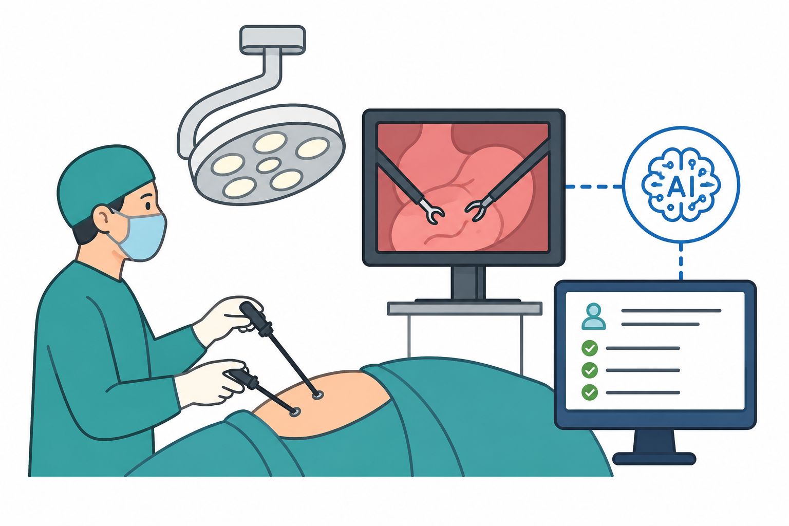
A groundbreaking artificial intelligence (AI) tool has been developed at The University of Texas Health Science Center at San Antonio, enabling rapid and accurate counting of brain lesions on MRI scans. Once integrated into clinical practice, this AI tool has the potential to aid neuroradiologists in the early evaluation of brain diseases in patients.
According to Dr. Mohamad Habes, a researcher at the Glenn Biggs Institute for Alzheimer’s and Neurodegenerative Diseases and Assistant Professor of Radiology, certain types of brain lesions are exceptionally challenging to quantify without AI.
In a study published in JAMA Network Open on April 24, Dr. Habes and his colleagues from multiple institutions demonstrated the utility of this AI tool in identifying and counting enlarged perivascular spaces (ePVSs). These spaces, which contain cerebrospinal fluid, surround arteries and veins, and serve as markers of cerebral small-vessel disease, a condition associated with stroke and dementia. The study analyzed data from 1,026 participants in the Multi-Ethnic Study of Atherosclerosis (MESA).
Dr. Habes emphasized the innovative nature of their deep-learning tool, which enables precise quantification of each individual ePVS in the brain, ultimately providing a comprehensive map of the patient’s small-vessel disease. Previously, the difficulty of manually counting ePVSs on MRI scans led to their disregard in clinical practice.
“On average, a middle-aged person may have around 500 or 600 of these small spaces on an MRI,” explained Dr. Habes. “Imagine a neuroradiologist attempting to manually count all of them. It simply isn’t feasible in a busy clinical setting, requiring one to two hours or more per scan.”
To address this challenge, the team developed an automated deep-learning algorithm for ePVS detection, as described in their publication in Neuroimage: Reports on March 7. By training the algorithm with expert knowledge, it is now capable of autonomously quantifying these lesions. The AI tool not only identifies the precise location of each ePVS but also counts them, measures their volumes, and provides a wealth of information that surpasses human capabilities.
This AI tool has the potential to revolutionize the evaluation of brain diseases, enabling quicker and more accurate diagnoses, and improving patient outcomes.
- Eurekalert